
Figure 2.
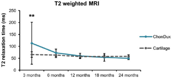
T2-weighted magnetic resonance imaging analysis of remodeled tissue within ChonDux-treated defects compared with adjacent uninjured cartilage. T2 relaxation times for adjacent uninjured cartilage were pooled from all patients (mean ± SD). Two-way analysis of variance with post hoc Tukey testing was performed to compare ChonDux time points and to uninjured cartilage. **P < 0.01 for ChonDux versus cartilage at 3 months.