. 2020 Jun 18;31(2):246–254. doi: 10.5606/ehc.2020.74405

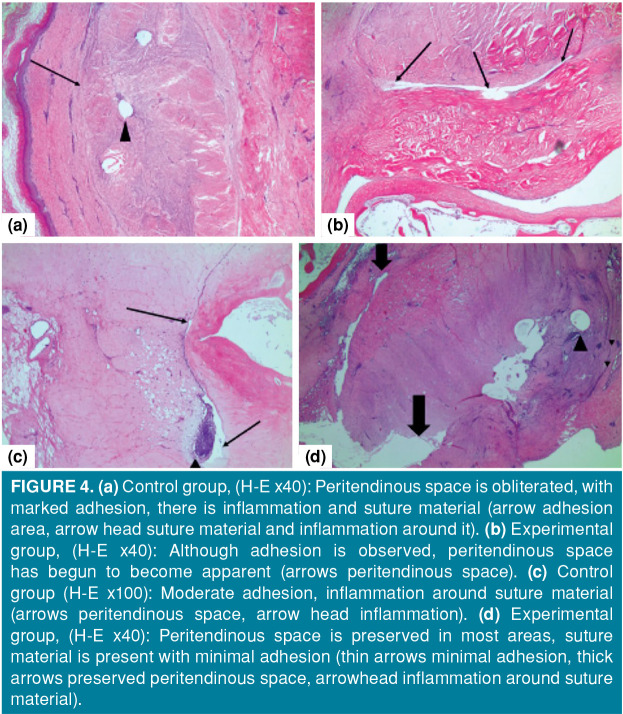
Figure 4. (a) Control group, (H-E x40): Peritendinous space is obliterated, with marked adhesion, there is inflammation and suture material (arrow adhesion area, arrow head suture material and inflammation around it). (b) Experimental group, (H-E x40): Although adhesion is observed, peritendinous space has begun to become apparent (arrows peritendinous space). (c) Control group (H-E x100): Moderate adhesion, inflammation around suture material (arrows peritendinous space, arrow head inflammation). (d) Experimental group, (H-E x40): Peritendinous space is preserved in most areas, suture material is present with minimal adhesion (thin arrows minimal adhesion, thick arrows preserved peritendinous space, arrowhead inflammation around suture material).

Figure 4