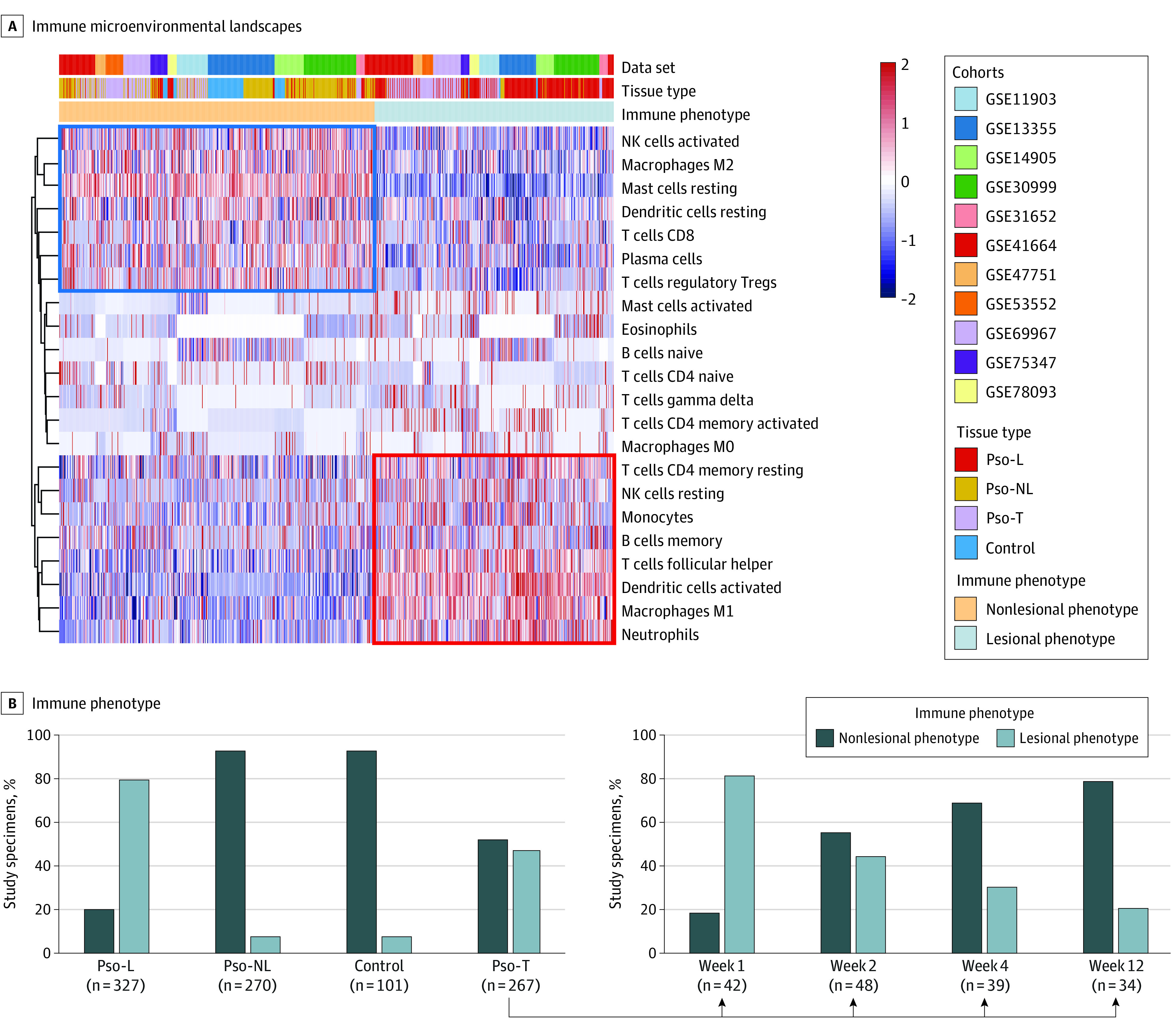
Figure 1. Distinct Immune Microenvironment Landscapes Used to Define Psoriasis Plaques and Their Resolution.

Pso-L indicates psoriasis lesional; Pso-NL, psoriasis nonlesional; Pso-T, psoriasis treatment specimens. A, Vertical lines represent signal strength for each cell type depicted in rows. Shown is unsupervised clustering of immune cell signal strength from 1145 skin samples in multiple cohorts (GSE11903, GSE13355, GSE14905, GSE30999, GSE31652, GSE41664, GSE47751,GSE53552, GSE69967, GSE75343, and GSE78097) each measuring biopsies from Pso-L, Pso-NL, Pso-T, and control specimens. Nonlesional phenotype is more associated with healthy tissue, whereas lesional phenotype is more associated with lesional tissue. B, Although, in aggregate, all psoriasis lesions under treatment show a roughly equal mix between nonlesional phenotype and lesional phenotype, nonlesional phenotype proportion rises with elapsed time after treatment initiation, whereas lesional phenotype decreases.
