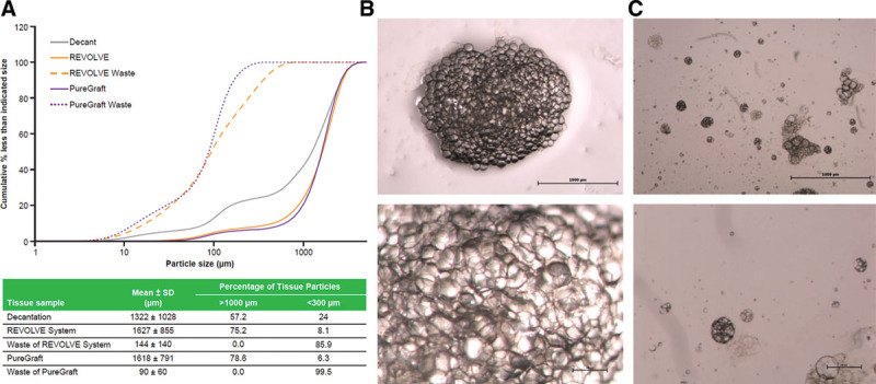
Fig. 1.

Fat tissue particle analysis. Fat graft was harvested after being processed with either decantation or the 2 filtration systems (REVOLVE System and PureGraft) and analyzed for fat particle size distributions and particle morphology as described in Materials and Methods section. A, Particle size distribution (accumulative frequency) from the different processed grafts indicated (upper). The data shown are from one representative lipoaspirate sample, and the characterization results for each processing method are listed in the table (lower). B and C, Images of fat tissues from the canister (B) and waste container (C) of the REVOLVE System. Upper row: 20×; lower row: 40×.
