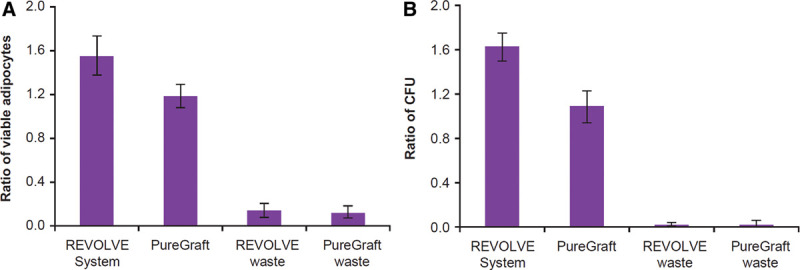
Fig. 4.

Evaluation of fat tissues harvested from the waste containers of filtration methods. Fat tissues filtered into the waste containers of 2 filtration methods were harvested, and both adipocytes and SVF cells were isolated for analysis as described in Materials and Methods section. A, Ratio of viable adipocytes in fat tissues when compared with the decantation graft (see the fat cell content in decantation sample in Fig. 2). B, Ratio of CFU count when compared with the decantation graft (see the number of CFU in the decantation sample in Fig. 3). CFU indicates colony-forming unit; SVF, stromal vascular fraction.
