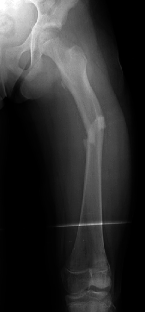
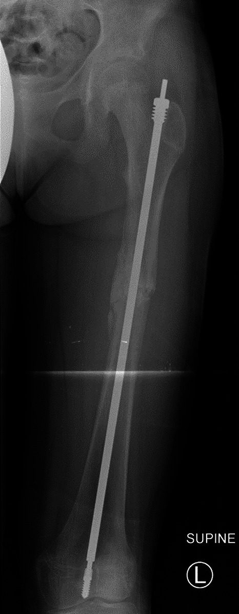
Figs. 1-A and 1-B Anteroposterior radiographs of the left femur of an 11-year-old girl with OI Type I.
Fig. 1-A.

Preoperative radiograph showing the left femur with a mid-diaphyseal femoral fracture sustained while the patient was dancing competitively.
Fig. 1-B.

Postoperative radiograph showing a 5.4-mm Fassier-Duval expanding rod.
