
Figs. 3-A and 3-B Anteroposterior radiographs of the lower limbs of a 27-month-old boy with OI Type III and severe deformity of the femora and tibiae. The patient has been undergoing cyclic bisphosphonate treatment since infancy; his weight was 9.5 kg.
Fig. 3-A.
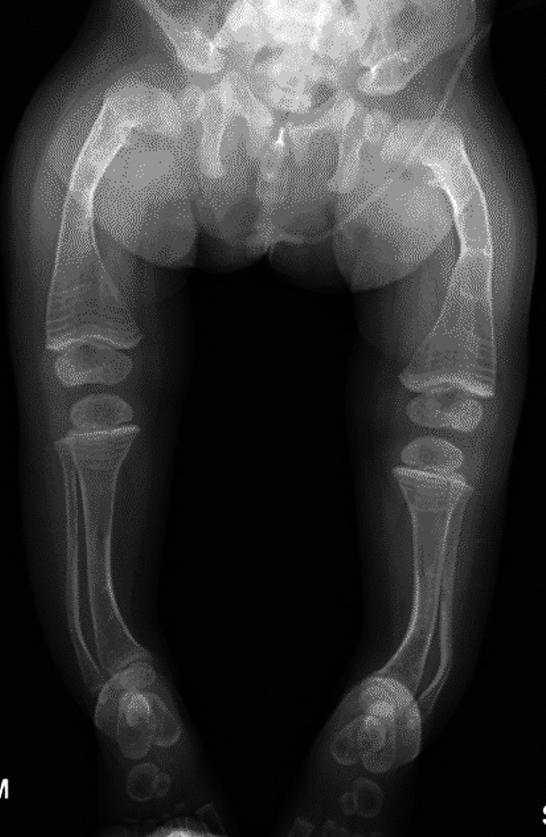
Preoperative radiograph showing the severe deformity of the femora and tibiae.
Fig. 3-B.

Postoperative radiograph. When the patient became more mobile and attempted pulling to stand, he underwent fragmentation and rodding of both femora and tibiae, the 2 legs (the tibia and the femur in each) were staged 2 weeks apart. The diameter of the tibiae precluded the use of expanding rods, so non-expanding rods were used in both tibiae (2.38-mm [3/32-inch] Steinmann pins) and expanding rods (3.2-mm Fassier-Duval) were inserted in the femora.