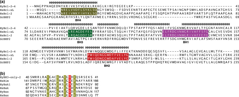
Figure 1. Sequence and structure alignment of hydra, human and sponge Bcl-2 proteins.
(a) Structure-based sequence alignment of HyBcl-2-4 with pro-survival Bcl-2 family members human Bcl-xL (PDB ID 4QNQ), human Bcl-2 (PDB ID 6G18) and G. cydonium BHP2 (PDB ID 5TWA). H denotes helical secondary structure based on HyBcl-2-4. The extent of the BH motifs are marked in color for hsBcl-xL and hsBcl-2 based on those defined in [5]; BH4, khaki; BH3, green; BH2, red; BH1, magenta. The structure-based sequence alignment was performed using Dali [20]. (b) Sequence alignment of BH3 motifs of Hy-BH3-only-2 (Genebank hma2.221399), HyBak1 (Uniprot T2MD83) and HyBax (Uniprot T2MDZ0) with human pro-apoptotic Bcl-2 proteins Bim (Uniprot O43521), Bak (Uniprot Q16611) and Bax (Uniprot Q07812). The four conserved hydrophobic residues are shaded in khaki, and the conserved aspartic acid in red. Sequence position numbers are given for each sequence.

