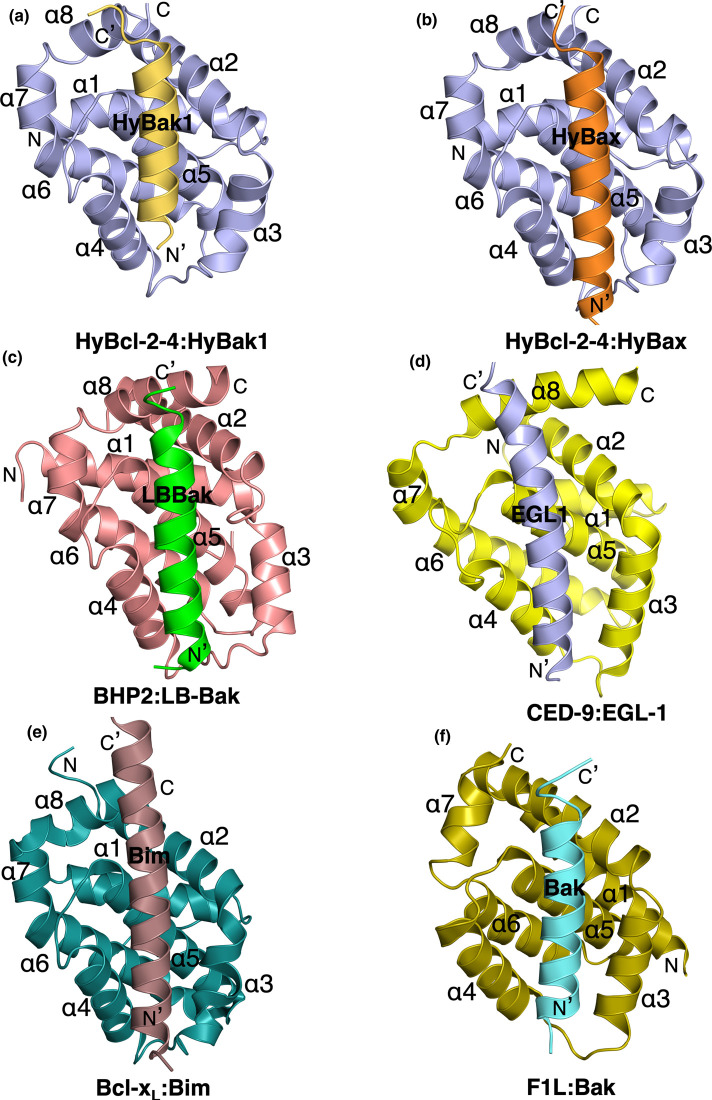
Figure 3. Ribbon representation of HyBcl-2-4 bound to HyBak1 BH3 and HyBax BH3 domain.
(a) HyBcl-2-4 (slate) in complex with the HyBak1 BH3 domain (sand). HyBcl-2-4 helices are labeled α1-α8. The view in (a) is into the hydrophobic binding groove formed by helices α2–α5. The view is directed towards the hydrophobic binding groove formed by helices α2–α5. (b) HyBcl-2-4 (slate) in complex with the HyBax BH3 domain (orange). HyBcl-2-4 helices are labeled α1–α8. (c) BHP2 (pink) in complex with the LB-Bak-2 BH3 domain (green) [12] (PDB ID 5TWA). (d) CED9 (yellow) in complex with the EGL1 BH3 domain (light blue) (PDB ID 1TY4) [34]. The view is as in (a). (e) Bcl-xL (teal) in complex with the Bim BH3 domain (chocolate) (PDB ID 1PQ1) [33] (a). (f) Vaccinia virus F1L (olive) in complex with the Bak BH3 domain (cyan) (PDB ID 4D2L) [35]. All views are as in (a).

