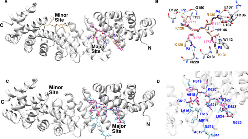
Figure 7. Comparison between ChREBP NLS and importin α interaction with those of SV-40 and AR.
(A) The superposed structure of importin α bound with ChREBP NLS 158–190 (pink) and SV-40 (tan, 1EJL). (B) Hydrogen bonding interactions of ChREBP (pink) and SV-40 (tan) at the P2–P5 pockets of major binding site of importin α. (C) Superimposed structures of AR (blue) and ChREBP NLS158–190 (pink) bound with importin α. (D) Close-up view of the superimposed structures of AR (blue) and ChREBP NLS 158–190 (pink) in the major site of importin α.

