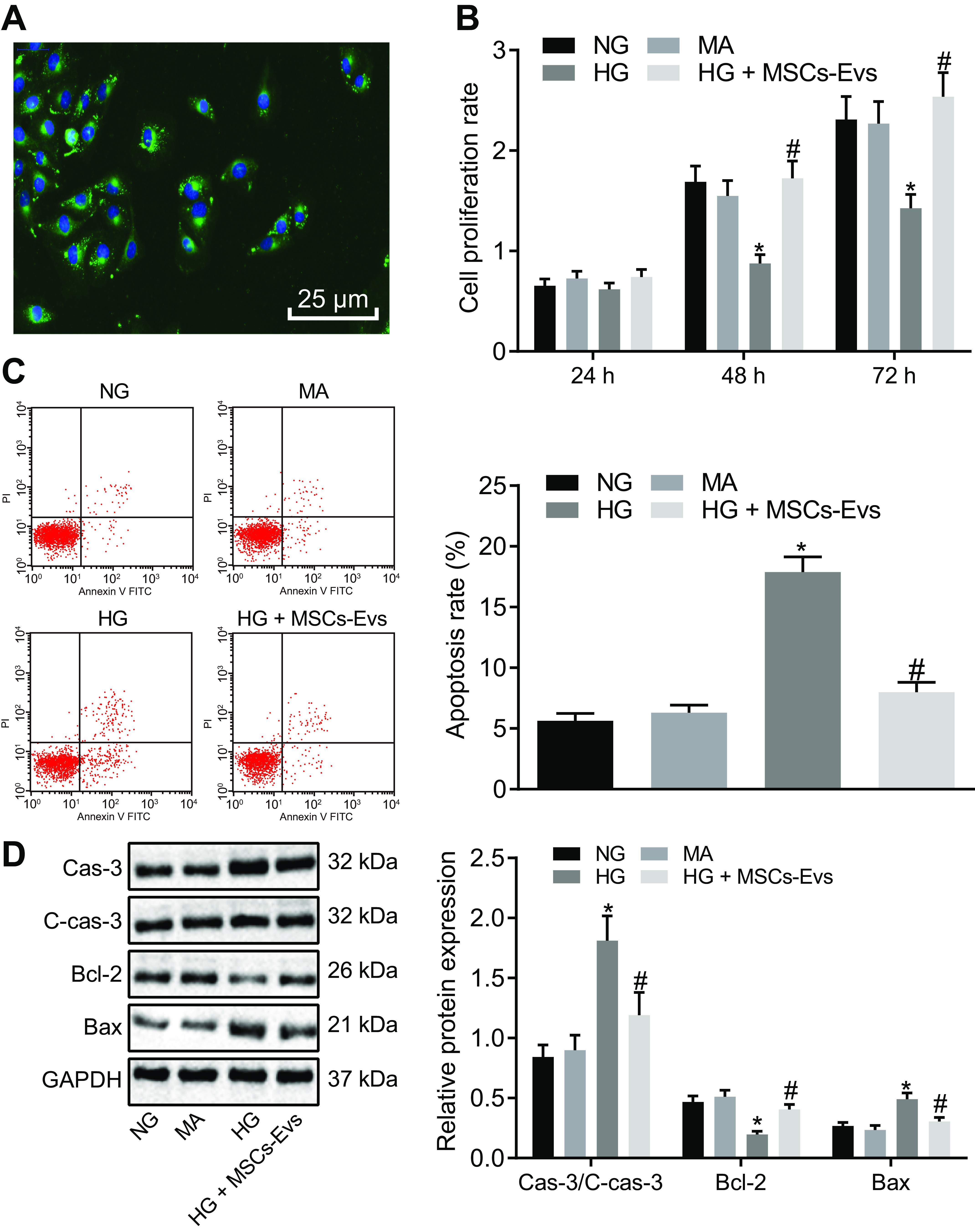
Figure 4.

ADSC-derived EVs inhibit apoptosis of MP5 cells induced by HG. A, the PKH67-labeled ADSC-derived EVs observed under a fluorescence microscope (green fluorescence, PKH67; blue fluorescence, 4′,6-diamidino-2-phenylindole) (scale bar, 25 μm). B, viability of MP5 cells after 24 h, 48 h, and 72 h of treatment as detected by CCK-8 assay. C, apoptosis of MP5 cells after treatment at 48 h, as detected by flow cytometry. D, the protein expression of apoptosis-related proteins (caspase-3, cleaved caspase-3, Bcl-2, Bax) in MP5 cells as detected by Western blotting analysis, normalized to GAPDH. Data are expressed as mean ± standard deviation. B–D, data among multiple groups were compared using one-way ANOVA, followed by Tukey's post hoc test. A, repeated-measures ANOVA was used to compare data among multiple groups at different time points, followed by Bonferroni post hoc test. The experiment was repeated three times. *, p < 0.05 versus NG or MA. #, p < 0.05 versus HG.
