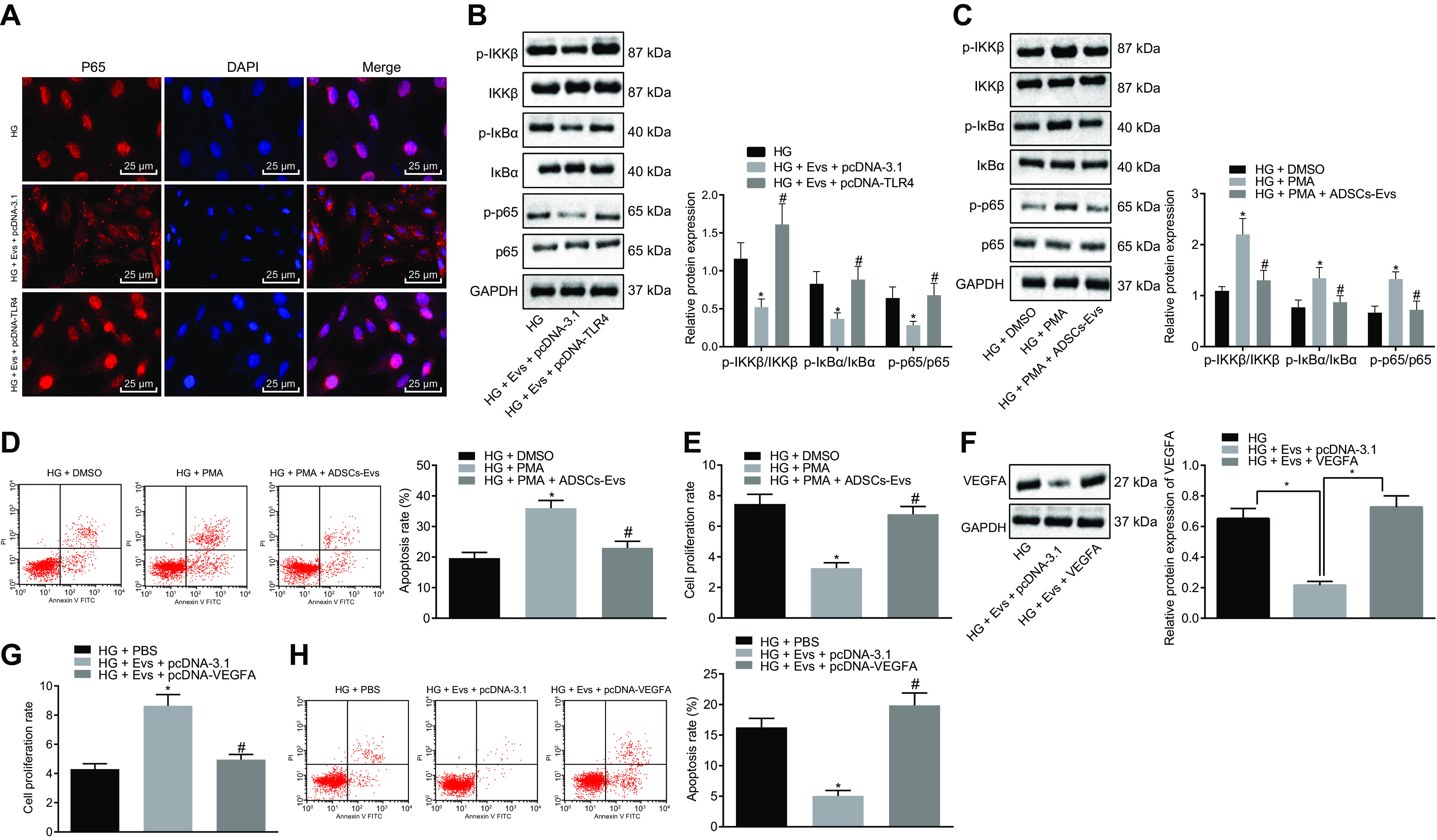
Figure 7.

EVs derived from ADSCs deliver MiR-26a-5p, which targets TLR4, downregulates NF-κB/VEGFA, and inhibits the apoptosis of MP5 cells induced by HG. A, immunofluorescence staining of p65 protein in MP5 cells (scale bar, 25 μm). B, the protein expression of NF-κB pathway-related proteins IKKβ, IκBα, and p65 as well as the extent of IKKβ, IκBα, and p65 phosphorylation in MP5 cells as detected by Western blot analysis, normalized to GAPDH. *, p < 0.05 versus HG. #, p < 0.05 versus HG + pcDNA-TLR4 + Exo. C, the protein expression of the NF-κB pathway-related proteins IKKβ, IκBα, p65, and VEGFA as well as the extent of IKKβ, IκBα, and p65 phosphorylation in HG-induced MP5 cells treated with ADSC-derived EVs and the NF-κB pathway activator, PMA, as detected by Western blot analysis, normalized to GAPDH. D, the apoptosis of HG-induced MP5 cells treated with ADSC-derived EVs and the NF-κB pathway activator, PMA, as detected by flow cytometry. E, cell viability in HG-induced MP5 cells treated with ADSC-derived EVs and the NF-κB pathway activator, PMA, as detected by CCK-8 assay. *, p < 0.05 versus HG + DMSO. #, p < 0.05 versus HG + PMA. F, the protein expression of VEGFA in HG-induced MP5 cells after treatment with ADSC-derived EVs and overexpression of VEGFA as detected by Western blot analysis, normalized to GAPDH. *, p < 0.05 versus HG + Exo + pcDNA-3.1. G, cell viability in HG-induced MP5 cells after treatment with ADSC-derived EVs overexpressing VEGFA, as detected by CCK-8 assay. H, cell apoptosis in HG-induced MP5 cells after treatment with ADSC-derived EVs overexpressing VEGFA as detected by flow cytometry. Data are expressed as mean ± standard deviation. Data among multiple groups were compared using one-way ANOVA, followed by Tukey's post hoc test. The experiment was repeated three times. *, p < 0.05 versus HG. #, p < 0.05 versus HG + Exo + pcDNA-3.1.
