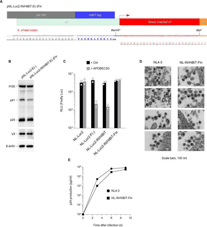
Figure 2.
Rescued Vif activity results in intact anti-APOBEC3G activity and replication ability. A, reconstruction of the HiBiT-tagged HIV-1 proviral DNA that rescues Vif function. After mutating the vif initiation codon (indicated by the red arrowhead), the fragment encoding the 25 amino acid sequence (shown in red) was inserted immediately downstream of the HiBiT tag into BamHI and BlpI sites, the latter of which was newly introduced at the N terminus of the Vif sequence (both sites with asterisks were disrupted by inserting the fragment). B, anti-p24 (upper), anti-Vif (middle), and anti–β-actin (lower) immunoblots of cell extracts transfected with either pNL-Luc2-E(−) or pNL-Luc2-IN/HiBiT-E(−)Fin. Data shown are representative of two independent experiments. C, infection of HeLa cells by VSV-G-pseudotyped luc-reporter WT (NL-Luc2), Vif-deficient NL-Luc2 (NL-Luc2-F(−)), HiBiT-tagged NL-Luc2 (NL-Luc2-IN/HiBiT), and HiBiT-tagged/Vif-inserted NL-Luc2 (NL-Luc2-IN/HiBiT-Fin), produced from cells expressing a vector control (black) or APOBEC3G (gray). Data from two experiments are shown (mean ± S.D., n = 3 technical replicates). RLU, relative light units. D, a representative transmission EM image of NL4-3 and NL-IN/HiBiT-Fin virions accumulated at the surface. Bars, 0.1 μm. E, multiple rounds of virus replication in peripheral blood mononuclear cells. PHA-IL-2-stimulated cells (2.5 × 10⁵) were infected with 25 ng of p24 antigen of either the NL4–3 (filled circle) or NL-IN/HiBiT-Fin (filled square) virus. Supernatants were harvested at the indicated times, and virus replication was monitored using p24 ELISA. The data shown are representative of three independent experiments.

