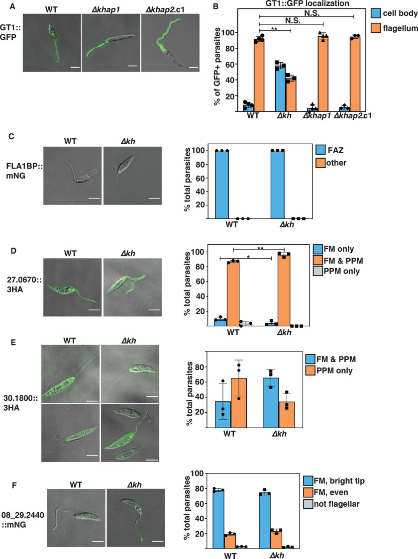
Figure 7.
KHAP1, KHAP2, and KH are not involved in targeting of various proteins to the flagellar membrane. A and B, KHAP1 and KHAP2 are not required for GT1::GFP trafficking to the flagellar membrane. A, live cell imaging of WT (left panel), Δkhap1 (middle panel), and Δkhap2.c1 (right panel) promastigotes episomally expressing GT1::GFP, imaged for GFP fluorescence and DIC in CyGEL. B, quantification of GT1::GFP localization, with cells categorized by the localization of the bulk of fluorescent signal by an observer blind to the genetic strain. Data represent the average and S.D. of three independent experiments, with values for each plotted on the bar graph. For each sample, between 25 and 40 cells were counted for each independent experiment. C–F, four other membrane proteins do not depend on KH for flagellar targeting. C, FLA1-binding protein LmxM.10.0620, FLA1BP; D, putative amino acid permease LmxM.27.0670; E, putative amino acid permease LmxM.30.1800; F, putative cyclic nucleoside monophosphate phosphodiesterase LmxM.08_29.2440. For C and F, proteins were endogenously tagged at the C terminus with mNG, whereas for D and E, proteins were tagged at the C terminus with the 3HA tag and expressed from an episome. Panels at the left represent endogenous fluorescence (mNG) or immunofluorescence (3HA) for WT (WT) parasites, and panels on the right are similar images for Δkh null mutant lines. Graphs represent quantification of the % total cells with the indicated distribution of protein signals, for WT or Δkh parasites, as determined by microscopic examination between 48 and 428 parasites per sample in three replicate experiments. Error bars represent the standard deviation of the data, with triplicate values for each condition plotted on the bar graph. Statistical significance was determined between pairs using Student's t test, with N.S. indicating not significant, *, p < 0.05; **, p < 0.01. For C-F, all comparisons not marked by asterisks were not significantly different. Scale bars represent 5 μm. Abbreviations used in panels at the right are: FAZ, flagellum attachment zone; FM, flagellar membrane; PPM, pellicular plasma membrane.

