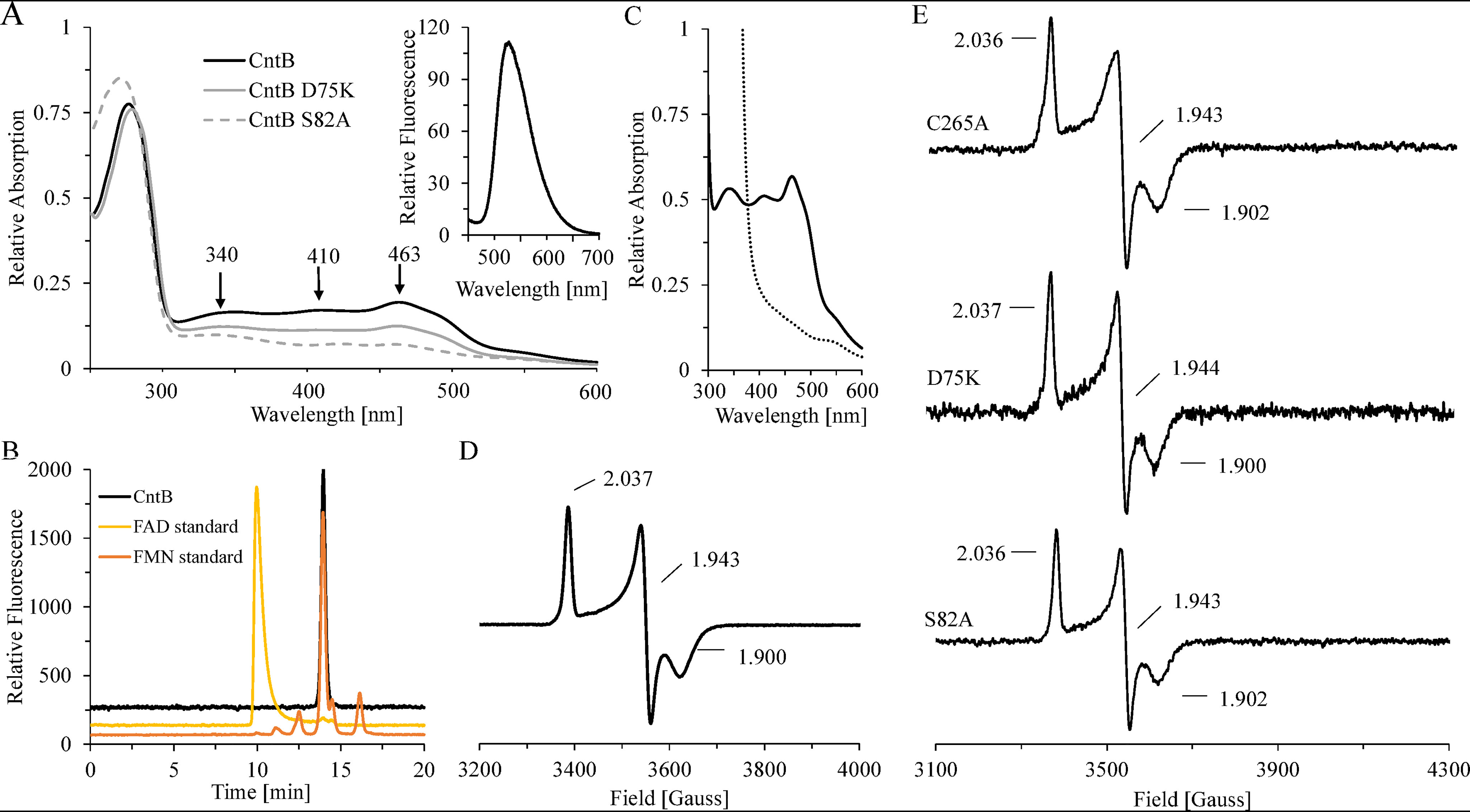
Figure 4.

Spectroscopic characterization of CntB WT and variant proteins. A, room temperature UV-visible spectra of purified CntB (black line) and variants D75K (gray line) and S82A (gray dashed line). Inset, fluorescence spectrum of a CntB supernatant after heat denaturation and centrifugation (370 nm excitation). B, identification of the cofactor FMN by HPLC. The fluorescent sample from panel A was chromatographed on a Reprosil 100 C18 column using excitation/emission wavelengths of 370/526 nm (black). Authentic samples of FAD (yellow) or FMN (orange) were analyzed accordingly. C, room temperature UV-visible spectrum of purified CntB before (black continuous trace) and after (black dashed trace) treatment with 2 mm dithionite for 10 min. D and E, low-temperature (15 K) EPR spectra of ∼1 mm CntB samples (WT or C267A, D75K, and S82A mutants) after dithionite reduction (10 mm). All spectra were recorded at 9.653 GHz, 7.5-G modulation amplitude, 0.2-milliwatt microwave power, and 100-kHz modulation frequency.
