
Figure 8.
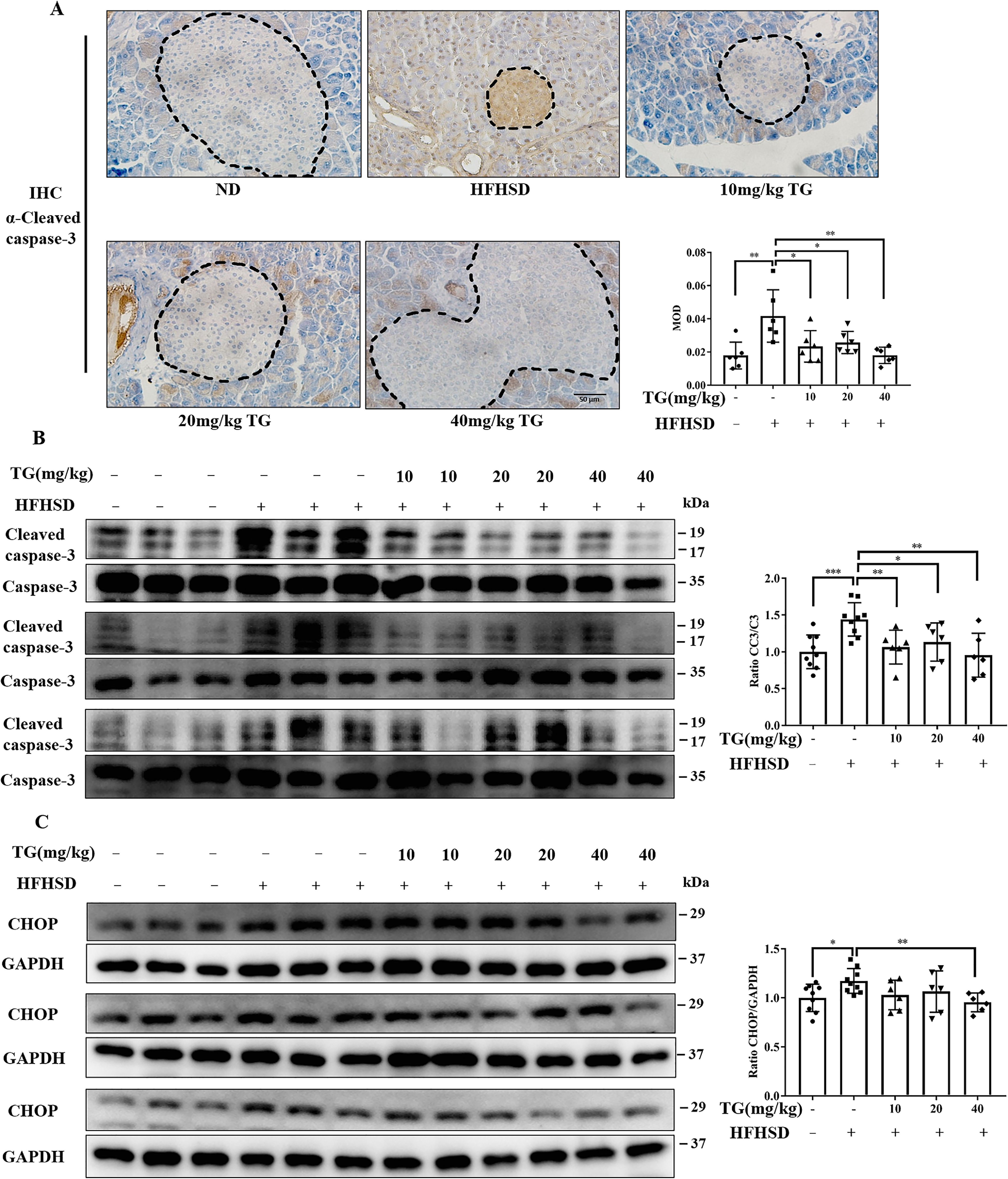
Therapeutic use of TG reduces apoptosis of islet β-cells by alleviating ER stress in HFHSD-induced diabetic mice. A, representative images of CC3 immunohistochemical staining of pancreas tissues (scale bar: 50 μm). Dashed lines and circled are islets. MOD of CC3, which was quantified by IPP software is presented in the bar graph. B and C, the protein levels of CC3 (B) and CHOP (C) were analyzed by Western blotting. Image J software was used for quantitative analysis. Error bars represent ±S.D. *, p < 0.05 normal control versus the HFHSD group; ‡, p < 0.05, 10 mg/kg of TG versus the HFHSD group; †, p < 0.05, 20 mg/kg of TG versus the HFHSD group; #, p < 0.05, 40 mg/kg of TG versus the HFHSD group; n ≥ 6.