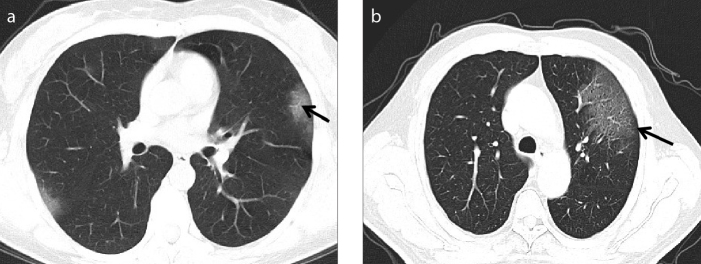
Figure 2. a, b.
Early stage CT findings of COVID-19. Axial CT image (a) of a 46-year-old female presenting with fever for 3 days. Flake-like ground-glass opacities are observed in the subpleural areas of upper lobes of bilateral lungs, in which thickened small vessels (black arrows) are seen. Axial CT image (b) of a 34-year-old male presenting with fever and cough for 6 days. A large sheet-like ground-glass opacity is seen in the upper lobe of left lung, in which the grid opacities are observed with the slabstone sign (black arrows).

