Figure 2.
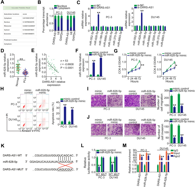
DARS-AS1 acts as a miR-628-5p sponge in PCa cells. (A) lncLocator predicted the cytoplasmic localization of DARS-AS1. (B) Relative amounts of DARS-AS1 in the cytoplasm and nucleus of PC-3 and DU145 cells were analyzed via RT-qPCR. (C) RT-qPCR was performed to detect miRNA (miR-552-3p, miR-628-5p, miR-188-5p, miR-6866-3p, miR-3200-5p, miR-370-3p, and miR-6893-3p) expression in PC-3 and DU145 cells after si-DARS-AS1 or si-NC transfection. (D) The expression level of miR-628-5p was detected by RT-qPCR in 53 pairs of PCa tissues and adjacent normal tissues. (E) Pearson’s correlation coefficient was applied to determine the correlation between the levels of DARS-AS1 and miR-628-5p in PCa tissues. (F) miR-628-5p expression in miR-628-5p mimic-transfected or mimic control-transfected PC-3 and DU145 cells was measured by RT-qPCR analysis. (G, H) CCK-8 assay and flow cytometry analysis were performed to evaluate the proliferation and apoptosis of miR-628-5p-overexpressing PC-3 and DU145 cells. (I, J) The impacts of miR-628-5p upregulation on the migration and invasion of PC-3 and DU145 cells were assessed by Transwell assays. (K) The predicted binding site between DARS-AS1 and miR-628-5p. The mutant sequences were also shown. (L) Luciferase reporter assays were performed in PC-3 and DU145 cells transfected with miR-628-5p mimic or mimic control to detect the luciferase activity of reporter DARS-AS1-WT and DARS-AS1-MUT. (M) The enrichment of miR-628-5p and DARS-AS1 in Ago2-containing immune precipitated RNA was quantified by RT-qPCR. *P < 0.05 and **P < 0.01.

