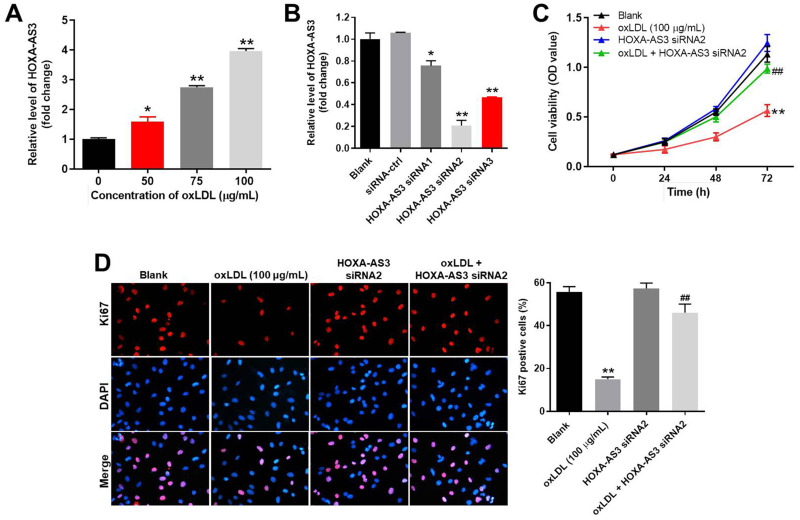
Figure 1.
OxLDL-induced growth inhibition of HUVECs was significantly reversed by HOXA-AS3 knockdown. (A) HUVECs were treated with 0, 50, 75 or 100 μg/mL oxLDL for 48 h. Then, the expression of HOXA-AS3 in HUVECs was detected by RT-qPCR. (B) HUVECs were transfected with HOXA-AS3 siRNA1, siRNA2 or siRNA3 for 24 h. Then, the expression of HOXA-AS3 in HUVECs was measured by RT-qPCR. (C) HUVECs were treated with oxLDL (100 μg/mL), HOXA-AS3 siRNA2 or oxLDL + HOXA-AS3 siRNA2 for 0, 24, 48 or 72 h. The viability of HUVECs was detected by CCK-8 assay. (D) The proliferation of HUVECs was detected by Ki67 staining. The Ki67 positive rate was calculated. Red immunofluorescence indicated Ki67. Blue immunofluorescence indicated DAPI. *P< 0.05 compared to control, **P< 0.01 compared to control, ##P< 0.01 compared to oxLDL (100 μg/mL); n = 3.

