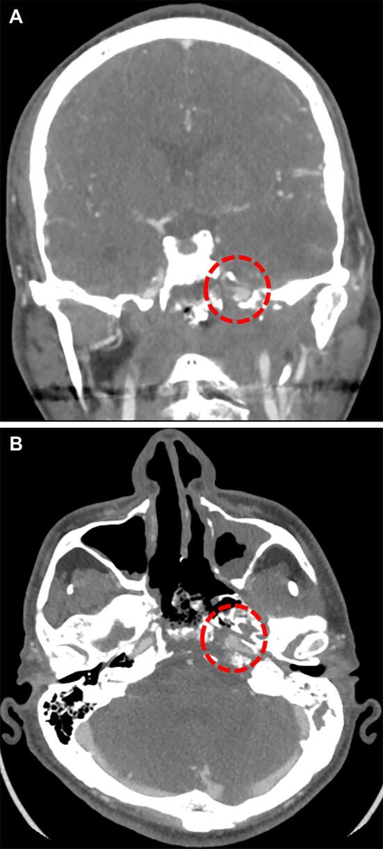
Figure 1.

CT-angiogram demonstrating findings of radiation-induced injury to the internal carotid artery. This is a patient with a history of nasopharyngeal carcinoma treated with chemoradiation at initial diagnosis and then recurrent, who subsequently presented with massive epistaxis. CT-angiography at the time demonstrated a pseudoaneurysm involving the horizontal petrous segment of the left internal carotid artery along with significant treatment-related changes in the adjacent bony skull base A and B. After passing a balloon test occlusion, the carotid artery was sacrificed.
