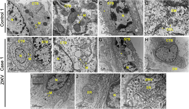
FIGURE 2.
Electron microscopy analysis of placental sections showed alterations and virus-like particles in ZIKV infected samples. (A–D) Electron microscopy images of ultrathin sections of placental tissue from a single, non-ZIKV infected mother that exhibited regular cytotrophoblasts (CTB), syncytiotrophoblasts (STB), nucleus (N), mitochondria (M), and collagen filament structures (COL). (E–H) Electron micrographs of ultrathin sections of placental tissue from different ZIKV-infected mothers showing CTB with alterations in the cytoplasm, nuclear variation (N) and swollen mitochondria with a loss of cristae and membrane rupture. The identified STB presented an enlargement of vesicles and apoptotic bodies (asterisks) along with an absence of normal membrane extensions and evidence of microvesicle secretion. The extracellular matrix (EM) did not present collagen filaments. (I–K) Identification of clusters of virus-like particles that were positioned near the endoplasmic reticulum (ER) of CTB and ∼25 nm in diameter, which is consistent with the dimensions of ZIKV.

