FIGURE 6.
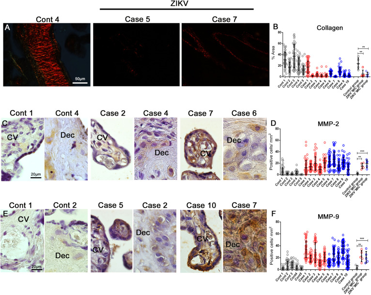
Detection and quantification of collagen, MMP-2 and MMP-9 collagenases expression. (A) Collagen detection by Picro Sirius Red staining in placental tissues. (B) The percent collagen area was quantified in all cases that showed a decrease in the expression of collagen in infected placentae. (C–E) Detection of MMP-2 and MMP-9 in decidual cells and cells located within the chorionic villi in both control and ZIKV infected placentae. (D–F) Quantification of the number of cells expressing MMP-2 and MMP-9 showed an increased expression in ZIKV infected placental tissues. Data are represented as mean ± SDM. Asterisks indicate differences that are statistically significant between groups (**p < 0.01) or (***p < 0.001).

