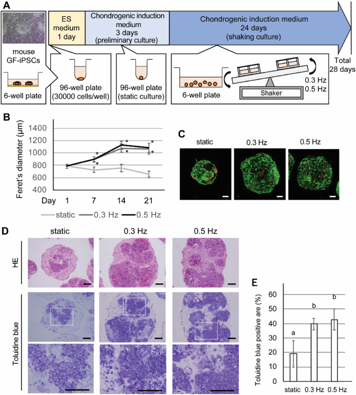
Figure 1.
Effects of a shaking culture on chondrogenically induced iPSC (CI-iPSC) construct aggregation profiles. (A) Fabrication of CI-iPSC constructs. (B) The size of 5 randomly selected CI-iPSC constructs was measured using ImageJ software on culture days 1, 7, 14 and 21. Experiments were repeated three times with similar results. Representative data from three independent experiments are shown (mean values ± SD: n = 5). Asterisks indicate statistically significant differences with respect to the static group (P < 0.01, Dunnett’s correction for multiple comparisons). (C) Representative fluorescence images of live/dead (green/red) cells at a depth of approximately 200 µm from the surface of the 21-day CI-iPSC constructs under the static and shaking conditions (0.3 and 0.5 Hz). Scale bars; 100 μm. (D) Representative histological image of HE staining and toluidine blue staining obtained from 21-day cultures under the static and shaking conditions (0.3 and 0.5 Hz). Scale bars; 100 μm. (E) Quantitative analysis of toluidine blue-positive area. Experiments were performed in quintuplicate (5 different constructs) and repeated three times with similar results. Representative data from three independent experiments are shown (mean values ± SD: n = 5). Different letters indicate significant differences between groups (P < 0.01, ANOVA with Tukey’s multiple comparison test).

