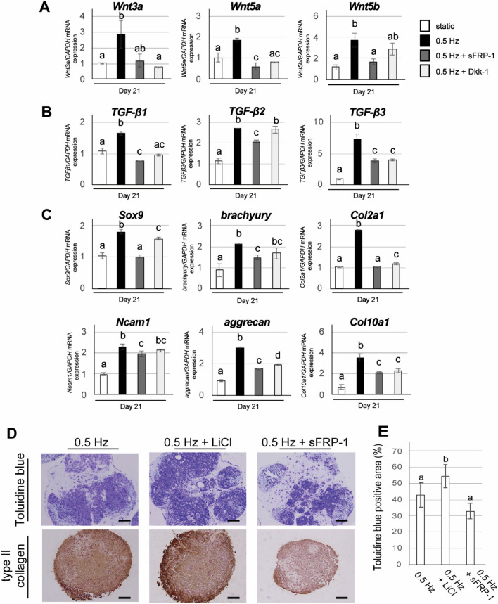
Figure 6.
Effects of Wnt signaling pathway inhibitors (sFRP-1 and Dkk-1) and activator (LiCl) on shaking culture-enhanced chondrogenic differentiation of iPSC constructs. (A–C) iPSCs were subjected to chondrogenic differentiation under static or shaking (0.5 Hz) culture in the presence or absence of sFRP-1 (100 ng/ml) or Dkk-1 (100 ng/ml). Gene expression of (A) Wnt3a, Wnt5a and Wnt5b, (B) TGF-β1, TGF-β2 and TGF-β3, (C) Sox9, brachyury, Col2a1, N-cam1, aggrecan and Col10a1 was determined by quantitative real-time RT-PCR at day 21. Experiments were performed in triplicate and repeated three times with similar results. Representative data from three independent experiments are shown (mean values ± SD: n = 3). Different letters indicate significant differences between groups (P < 0.05 for brachyury and Ncam1, and P < 0.01 for other genes, ANOVA with Tukey’s multiple comparison test). (D, E) iPSCs were subjected to chondrogenic differentiation under shaking (0.5 Hz) culture in the presence or absence of LiCl (5 mM) and sFRP-1 (100 ng/ml). (D) Representative histological images of CI-iPSC constructs at day 21, which were subjected to toluidine blue staining and immunohistochemical detection of type II collagen. Scale bars; 100 μm. (E) Quantitative analysis of toluidine blue-positive area. Experiments were performed in quintuplicate (5 different constructs) and repeated three times with similar results. Representative data from three independent experiments are shown (mean values ± SD: n = 5). Different letters indicate significant differences between groups (P < 0.05, ANOVA with Tukey’s multiple comparison test).

