
Fig. 6. Linsitinib with a suboptimal dose of osimertinib regresses AXL-low tumors in vivo.
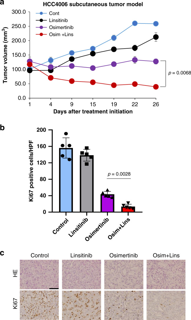
a HCC4006 cell line-derived xenograft (CDX) tumors were treated with vehicle (control:n = 4 mice), linsitinib 50 mg/kg (n = 4 mice), osimertinib 5 mg/kg (n = 5 mice), or linsitinib 50 mg/kg plus osimertinib 5 mg/kg (n = 5 mice), administered daily by oral gavage. Tumor volumes were measured over time from the start of treatment (mean ± s.e.m.). p values are provided (two-sided Student’s t-tests). b Quantification of proliferating cells, as determined by their Ki-67-positive proliferation index (percentage of Ki-67-positive cells) as described in “Methods”. Columns, mean of five areas. Data are presented as mean ± s.d. p values are provided (two-sided Student’s t-tests). HPF high power field. c Representative images of HCC4006 xenografts for H&E staining and immunohistochemical staining with antibodies to human Ki-67. Bar, 100 μm.