
Fig. 3.
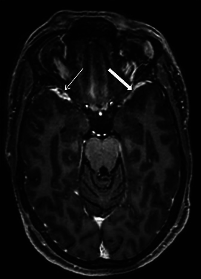
Postcontrast thin 3D T1 MPRAGE image showed nonocclusive filling defects suggestive of clots within the right sphenoparietal venous sinus at the anterior temporal pole (thin arrow). Note the normal contrast-filled left sphenoparietal sinus for comparison (thick arrow).