Fig. 1.
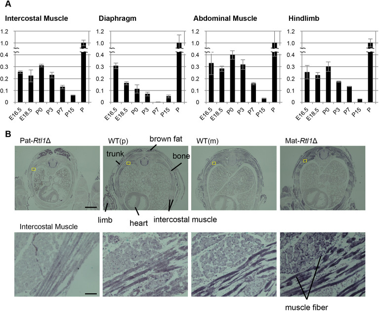
Rtl1 mRNA expression in embryos, and in embryonal and neonatal muscles; RTL1 protein localization in neonatal intercostal muscles. (A) Quantitative PCR results of Rtl1 in the diaphragm, hindlimb, intercostal and abdominal muscles in E16.5 and E18.5 embryos and neonates (P0, P3, P7 and P15). Relative expression levels of Rtl1 to Gapdh are shown. Placenta (P: E18.5) was used as the positive control and its Rtl1/Gapdh ratio was adjusted to 1. Data are mean±s.d. (B) Immunohistochemical staining of the RTL1 protein in neonates. A cross-sectional view of the neonates (top) and higher magnification views of the intercostal muscle (yellow boxes) are shown (bottom). The RTL1 signals (purple by BCIP/NBT staining) were observed around and along the muscle fibers. Scale bars: 1 mm (top) and 50 μm (bottom). Neonates were fixed in Super Fix.

