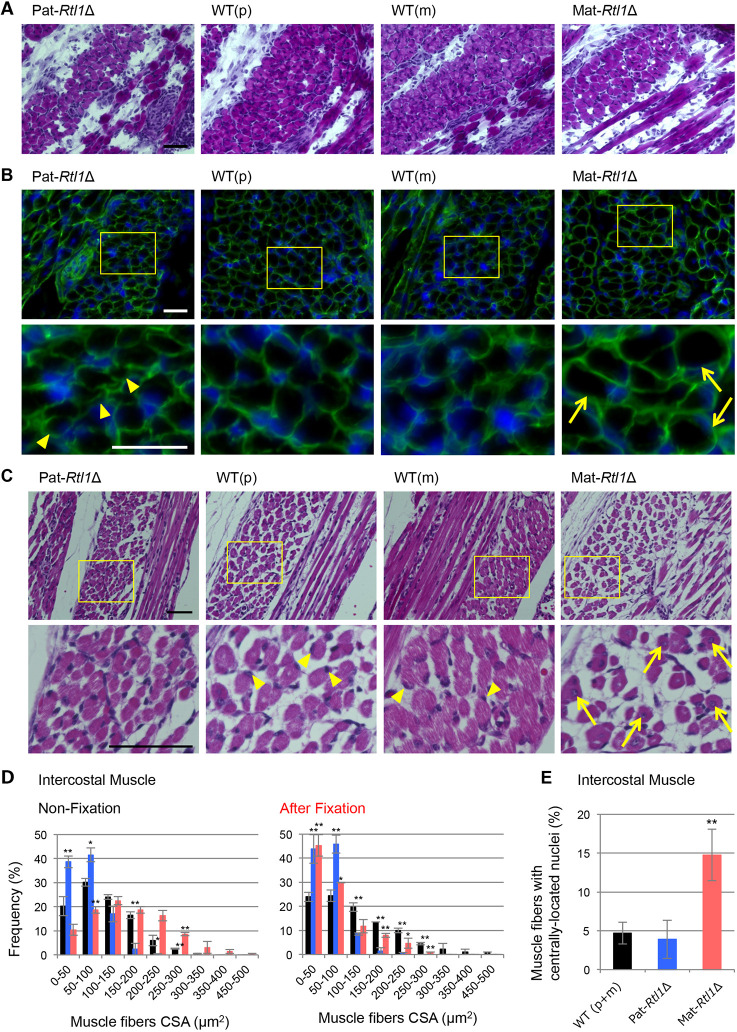
Fig. 2.
Histological abnormalities in intercostal muscle of Pat- and Mat-Rtl1Δ. (A,B) Hematoxylin and Eosin (HE) staining and immunofluorescence staining of the neonatal intercostal muscle. (A) HE staining of the neonatal intercostal muscle. (B) Co-immunostaining with laminin (green) and DAPI (blue) (top row), and higher magnification views of the intercostal muscle (yellow boxes) (bottom row). The arrowheads in the Pat-Rtl1Δ column indicate thinner muscle fibers and the arrows in the Mat-Rtl1Δ column indicate large muscle fibers. The neonates were not fixed before being embedded in OCT compound. (C) HE staining in neonate intercostal muscle (top) and higher magnification views (bottom): Pat-Rtl1Δ (left), wild type (middle) and Mat-Rtl1Δ (right). The arrowheads in the wild-type columns indicate normal nuclei and the arrows in the Mat-Rtl1Δ column indicate muscle fibers with centrally located nuclei. Scale bars: 50 μm. Neonates were fixed in Super Fix. (D) Distribution of the muscle fiber cross-sectional area (CSA) in wild-type (black, n=4), Pat-Rtl1Δ (blue, n=3) and Mat-Rtl1Δ (red, n=3) neonates [non-fixed samples (left) and fixed samples with Super Fix (right)]. (E) Proportion of muscle fibers with centrally located nuclei (arrows in C) between wild-type (black, n=4), Pat-Rtl1Δ (blue, n=4) and Mat-Rtl1Δ (red, n=4) neonates. Neonates were fixed in Super Fix. *P<0.05, **P<0.01 (two-tailed Student's t-test). Data are mean±s.d.

