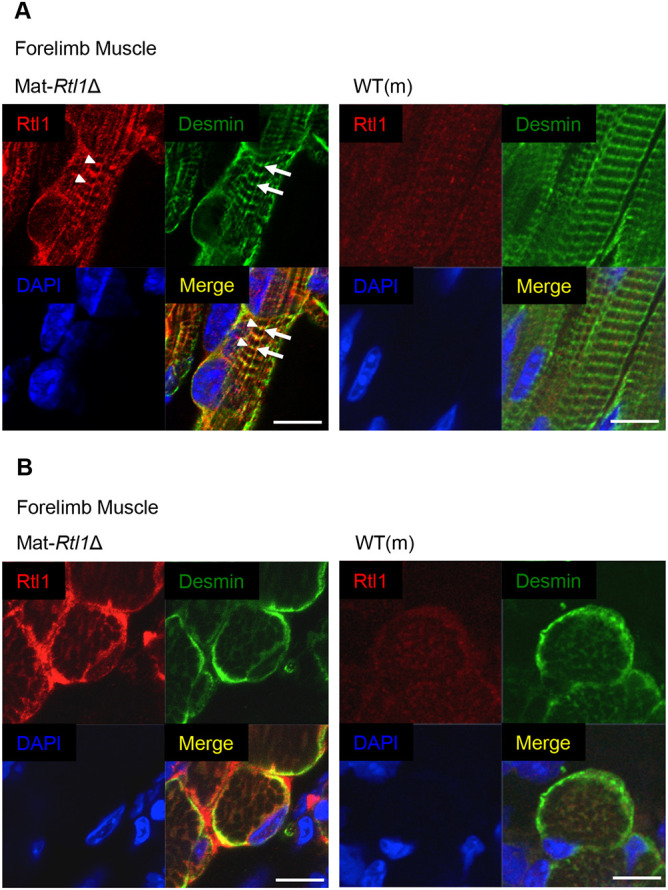
Fig. 3.

Expression of Rtl1 in the neonatal muscle. (A,B) Immunofluorescence staining of RTL1 protein in the neonatal forelimb muscles from Mat-Rtl1Δ and WT(m) mice. Long axis views (A) and cross-sectional views (B) of the muscle fibers. Co-immunostaining with RTL1 (red; arrowheads), desmin (green; arrows) and DAPI (blue), and their merged images. Scale bars: 20 μm. The neonates were not fixed before being embedded in the OCT compound.
