
Figure 2.
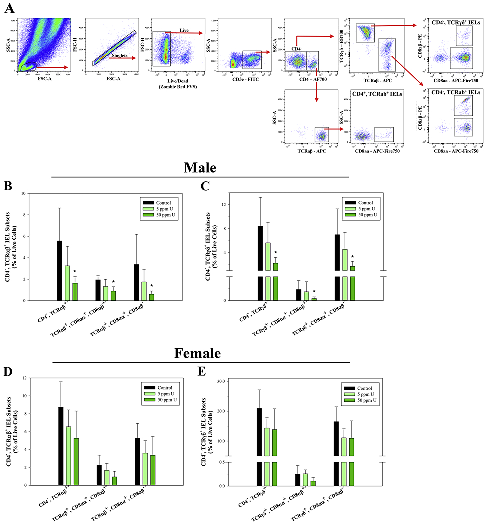
U exposure suppresses CD3ε+, CD4−, TCRαβ+ and TCRγδ+ IEL subsets in the small intestine of male mice. Small intestinal intraepithelial cells were isolated from male and female mice following 45-day exposure to 0, 5, and 50 ppm U and IEL subsets were evaluated based on surface marker phenotype by flow cytometry. (A) Representative flow cytometry plots demonstrating the gating strategy used to define small intestinal IEL subsets. Percentages of (B) CD3ε+, CD4−, TCRαβ+ and (D) CD3ε+, CD4−, TCRγδ+ IEL subsets in male mice. Percentages of (E) CD3ε+, CD4−, TCRαβ+ and (F) CD3ε+, CD4−, TCRγδ+ IEL subsets in female mice. Data are expressed as mean ± SD. Male and female: n = 6 mice/group. *p<0.05 in one-way ANOVA followed by a Dunnett’s t-test or Kruskal-Wallis One Way ANOVA on Ranks followed by Dunnett’s post-hoc test compared to control group. Abbreviations: FVS, fixable viability stain; AF700, Alexa Fluor 700.