
Figure 3.
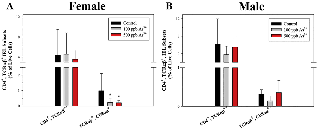
Small intestinal CD3ε+, CD4+, TCRαβ+, CD8αα+ IELs are reduced by As3+ exposure in male and female mice. Intraepithelial cells were isolated from the small intestines of male and female mice following 45-day exposure to 0, 100, and 500 ppb As3+ and IEL subsets were evaluated based on surface marker phenotype by flow cytometry. Percentages of CD3ε+, CD4+, TCRαβ+ IEL subsets in (A) female and (B) male mice. Data are expressed as mean ± SD. Male: control (n = 5), 100 ppb (n = 3), and 500 ppb As3+ (n = 5). Female: control (n = 4), 100 ppb (n = 6), and 500 ppb As3+ (n = 6). *p<0,05 in one-way ANOVA followed by a Dunnett’s t-test or Kruskal-Wallis One Way ANOVA on Ranks followed by Dunnett’s post-hoc test compared to control group.