
Fig. 13.6.
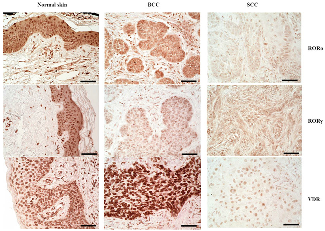
Immunohistochemical detection of RORα(upper), RORγ (middle), and VDR (lower) in normal skin (left panel), BCC (middle), and SCC (right). Scale bar: 50 μm. Archival formalin-fixed paraffin-embedded sections, after heat-induced antigen retrieval in Trisbased antigen unmasking solution (Vector Laboratories, Inc., Burlingame, CA) and endogenous peroxidase blocking, were incubated over night at 4 °C with primary antibodies (rabbit anti-RORα (provided by Dr. Anton M. Jetten), 1:400; rabbit anti-RORγ (provided by Dr. Anton M. Jetten), 1:50; rat anti-VDR (Abcam, MA1-710; Thermo Fisher Scientific, Waltham, MA)). Next, sections were incubated with secondary antibodies conjugated with HRP (anti-rabbit ImmPRESS antibody (ready to use, Vector Laboratories, Inc., Burlingame, CA) for RORα and RORγ; anti-rat antibody (1:200, Abcam, Cambridge, UK) for VDR), followed by peroxidase substrate ImmPACT NovaRED (Vector Laboratories Inc., Burlingame, CA, USA) application and mounting with permanent mounting media and glass coverslip (Thermo Fisher Scientific, Waltham, MA)