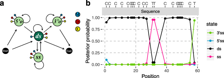
Fig. 2.
Graphical representation of the model (a) and illustration of the posterior decoding (b). a States are depicted by nodes and transitions by edges. Each state emits a match to the reference M (blue) or a mismatch, which can either be compatible with cytosine deamination, D (red), or an error (or polymorphism), E (yellow). Single-stranded states (5’o, 3’o and ss) and the double-stranded state (ds) are in light and dark green, respectively. b The posterior probability for each state is shown with different colours. (see Additional file 1: Supplementary Note 2 for an evaluation of the posterior decoding). We show bases on top for positions in the sequence (grey bar) that align to a C in the reference

