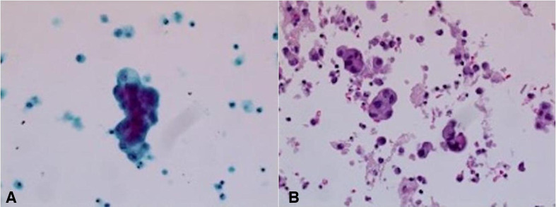
Figure 1.
(A) Thin prep, papanicolaou stain, ×400. (B) Cell block, H&E stain, ×200: the ascitic fluid in this patient shows three-dimensional clusters of epithelial cells with smooth community borders, raising the suspicion for adenocarcinoma. No windows are present. Background shows inflammatory cells.

