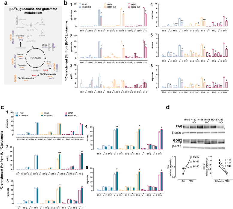
Fig. 3.
Dysregulation of glutamine and glutamate metabolism mirror each other in FTD3 neurons. a Scheme depicting the main labelling patterns from neuronal [U-13C] glutamine and [U-13C] glutamate metabolism and 13C-enrichment as percentage labelling in metabolites. Cultures were incubated for 90 min with b [U-13C] glutamine (0.5 mM), or c [U-13C] glutamate (0.25 mM) in the presence of unlabelled glucose (2.5 mM) then cell extracts were collected and analysed using GC-MS for determination of the percentage distribution of 13C-labelled metabolites in isogenic controls (pink bars/green bars) and the respective FTD3 patient cell lines incubated with 13C-labelled glutamine or glutamate (white bars). b The increased labelling in the TCA cycle metabolites and amino acids after [U-13C] glutamine incubation indicates increased metabolism of this amino acid. c In contrast, unmodified labelling in glutamate M + 5 and increased labelling in direct metabolites from [U-13C] glutamate indicates intact glutamate transport but decreased glutamate metabolism. d Protein expression assayed for by Western blot analyses of the two main enzymes involved in glutamine and glutamate degradation, namely PAG and GDH, respectively. Expression of PAG protein was upregulated while that of GDH was substantially reduced in FTD3 neurons from the three different patient cell lines (H150, H151 and H242). β-Actin was used as loading control. Results represent means ± S.E.M. obtained from three different patient cell lines indicated by small circles from three different culture preparations of hiPSC-derived neurons. *P < 0.05 or **P < 0.001, ***P < 0.0005, # P < 0.0001, two-way ANOVA correcting for multiple comparisons was employed

