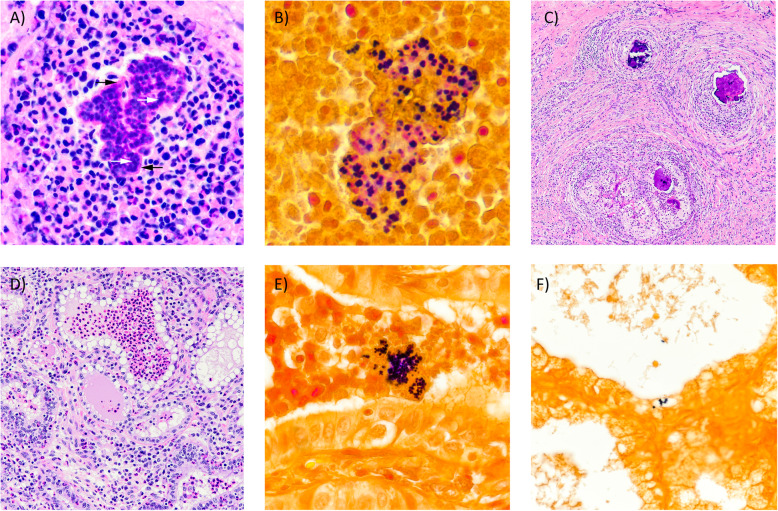
Fig. 3.
Photomicrographs of mastitic quarter from 1428. (A) Pyogranulomatous infiltrate. Note colonies of basophilic cocci (white arrows) within brightly eosinophilic and radiating matrix of Splendore-Hoeppli reaction (black arrows). H&E 20X. (B) Cocci within eosinophilic matrix are Gram-positive. Gram stain 40X. (C) Granulomas with abundant peripheral fibrosis and central areas of dystrophic mineralization. H&E 4X. (D) Acini containing numerous neutrophils. H&E 10X. (E) Intraluminal Gram-positive cocci within acinus. Gram stain 40X. (F) Intracellular Gram-positive cocci. Gram stain 20X

