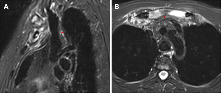
Fig. 3.
Soft tissue mass in the ACW region. a Sagittal water-only T2W Dixon image shows a soft tissue mass or infiltration around the hyperostotic bones in a 55-year-old female with a disease duration of 60 months. The lower part of the superior vena cava was normal, but the right brachiocephalic vein behind the soft tissue mass was slightly narrowed (red arrow). b Axial water-only T2W Dixon image reveals a soft tissue mass or infiltration behind the manubrium sterni in a 49-year-old male with a disease duration of 6 months. The left brachiocephalic vein was surrounded by the soft tissue mass and could not be evaluated (red arrow)

