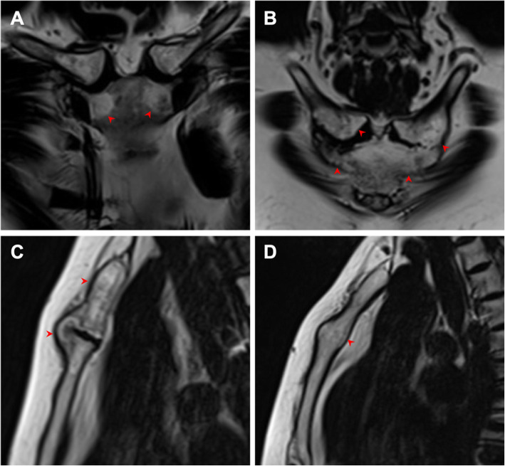
Fig. 5.
Chronic lesions in the ACW shown by oblique coronal (a–c) and sagittal (d, e) T2W Dixon images in patients with SAPHO syndrome. a Fat-only T2W Dixon image shows different shapes of fat infiltration, such as crescent in the corners of the manubrium, sheet in the right clavicle, and scattered lesions in the left clavicle. b Fat-only T2W Dixon image shows ossification of the costoclavicular ligament. c Fat-only T2W Dixon image shows a bone bridge of the sternal angle. d Fat-only T2W Dixon image shows hyperostosis in the sternal angle. Lesions are indicated by red arrows

