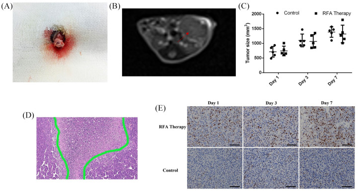
Figure 1.
The orthotopic PDAC murine models for RFA treatment. (A) Cell inoculation into the pancreas under surgery. (B) Follow-ups with MRI scanning in transverse plane; the tumor is marked with a red arrow. (C) Comparison of tumor volumes with or without RFA therapy on days 1, 3 and 7. (D) The typical necrotic area in the center of tumor post-RFA treatment. (E) Ki-67 staining of the residual tumor tissues with or without RFA therapy.
MRI, magnetic resonance imaging; PDAC, pancreatic ductal adenocarcinoma; RFA, radiofrequency ablation.

