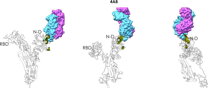
Figure 5.
Antibody 4A8–spike complex. The figure shows how 4A8 binds the N-domain of spike, supporting the correct prediction of the epitope. The MLCE-predicted epitope region is shown in green in three different orientations, indicating substantial contact formation with the antibody. The Fab of the antibody is depicted as accessible surface in shades of blue.

