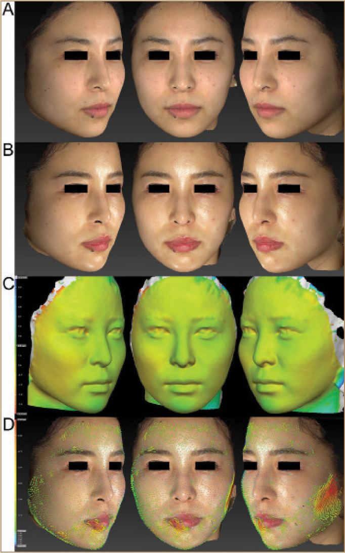
FIGURE 1.

A 22-year-old Japanese female; images from top to bottom show: A) pretreatment, B) six months posttreatment, C) superimposed 3D volumetric assessment comparing pre- and six months posttreatment, and D) quantification of 3D skin surface displacement with vectors comparing pre- and six months posttreatment. Marked improvements of skin texture and clarity were observed in 2D color digital photographs relative to pretreatment (A, B). Reductions in 3D volumetric assessment outcomes were observed when compared with pretreatment (C). The varying degrees of tightening achieved are shown in colors yellow to red (-5.0 mm). Green areas indicate areas what remained unchanged. Each point of the skin surface at the forehead, periorbital areas, and medial sides of the cheek was 3D displaced in a centripetal direction (D). The varying degrees of the 3D movement of the skin are shown in colors blue (0.3mm) to red (6.2mm). Green vectors indicate areas that were 3D displaced from 1.0mm to 2.5mm.
