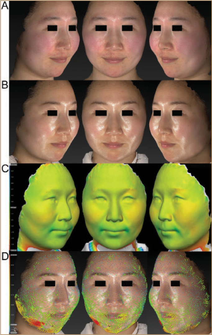
FIGURE 3.

A 27-year-old Japanese female; marked improvements in skin texture, redness, and clarity were observed on 2D color digital photographs relative to pretreatment (A, B). Tightening effects on the lower half of the face were observed during 3D volumetric assessment (C). Each point of the skin surface in the perinasal area and the cheeks was 3D displaced in a centripetal direction (D). The vectors in perioral area indicate downward. The varying degrees of the 3D movement of the skin are shown in colors blue (0.5mm) to red (9.1mm). Green vectors indicate areas that were 3D displaced from 1.4 to 3.6mm.
