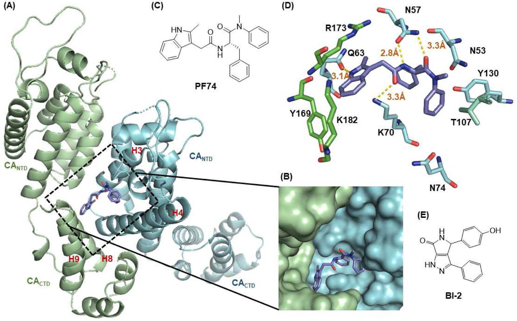
Figure 1.

Structure of HIV-1 CA and an important ligand binding pocket. (A) The highly helical structure of CA dimer (PDB: 4FXZ [28]). The pocket formed around H3 and H4 of the CANTD (cyan), and H8 and H9 of the adjacent CACTD (green) accommodates the binding of multiple ligands, including host factors Nup153 and CPSF6, and small molecules PF74 and BI-2; (B) close-up view of the pocket bound by PF74; (C) the chemical structure of PF74; (D) important residues providing key interactions with PF74; (E) the chemical structure of BI-2.
