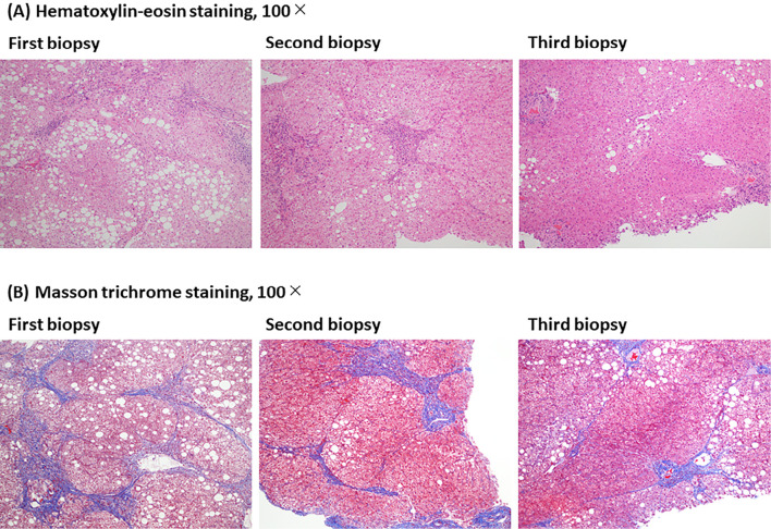
Figure 3.
Representative pathological images of an improved case (Case 4) at the third liver biopsy compared to pretreatment are shown. Histological changes at the three points of pretreatment (first liver biopsy), 24 weeks (second liver biopsy), and 3.5 years (third liver biopsy) after the start of SGLT2 inhibitor. (A) Hematoxylin and Eosin staining, 100×. (B) Masson trichrome staining, 100×.

