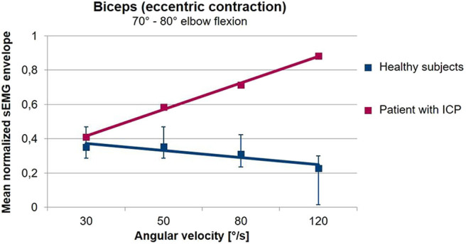
Figure 5.

Averaged sEMG envelopes of the Biceps Brachii as a function of the movement velocity during freely performed elbow extension movements. Values are normalized with respect to the 75% of the maximal value of the envelope. The sEMG when the elbow passes an interval from 80 to 70° flexion angle is analyzed. In healthy volunteers (blue), the biceps sEMG envelope decreases with increasing angular velocity. In contrast, in the patient with a spastic movement disorder (red) shown here, muscular activation increases with angular velocity. The gradient of the sEMG envelope–movement velocity relationship is thus a measure for the presence of spasticity during freely performed movements (unpublished data).
