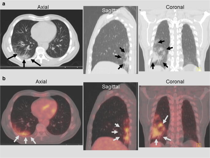
Fig. 1.
A 65-year-old male patient with locally advanced rectal cancer who came for [18F]-FDG PET/CT for initial staging. Axial, sagittal and coronal CT slices (a) and fused [18F]-FDG PET/CT (b) show the most frequent imaging finding in COVID-19 patients; this is GGOs (black arrows) coinciding with an area of intense [18F]-FDG uptake, peripherally localised, with an homogeneous [18F]-FDG uptake

