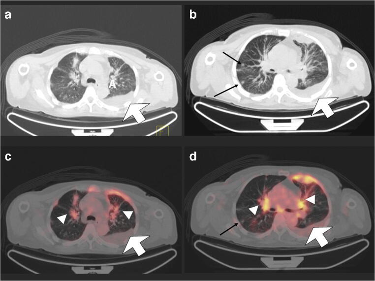
Fig. 3.
A 66-year-old female patient with head and neck tumour, referred for [18F]-FDG PET/CT for suspected recurrence. Axial CT (a and b) and fused [18F]-FDG PET/CT (c and d) show extensive disease in both lungs with pleural effusion (broad arrow), hilar infiltrates (arrow heads) and nodules (long arrows). GGOs were also present in this patient but are not included in the image presented

