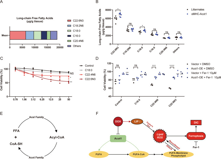
Fig. 7. αMHC-Acot1 transgenic mice show reshaped free fatty acid composition.
a The mean concentrations of free fatty acids detected by GC-MS in αMHC-Acot1 transgenic mice and their littermates. b Top 5 FFAs concentrations in murine heart tissue. c Cell viability analysis showing different concentrations of FFAs enhanced DIC in HL-1 cells. d Cell viability analysis showing the protective effect of Acot1 overexpression and Fer-1 co-treatment to FFAs enhanced DIC. e Diagrammatic method of showing the opposite enzymatic function of Acsl family and Acot family in the cell. f The potential mechanism of Acot1 inhibits the induction of ferroptosis in DOX-induced cardiotoxicity. LIP liable iron pool, DIC doxorubicin-induced cardiotoxicity, Fer-1 Ferrostatin-1. Significance in b was calculated using the unpaired Student’s t test. Significance in c, d was calculated using the one-way ANOVA test with multiple comparisons between the two groups. P value < 0.05 was considered to be significant, and labeled as *P < 0.05; **P < 0.01; ***P < 0.005; ****P < 0.001; ns not significant.

