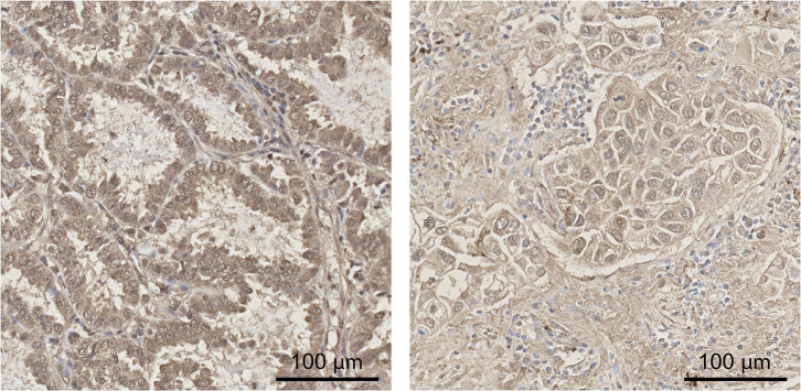
Figure 3.
Immunohistochemical staining of PAPP-A2. Expression of PAPP-A2 in lung cancer tissue was determined by immunohistochemical staining. Examples are shown for two patients with non-small cell lung cancer of adenocarcinoma subtype. PAPP-A2 staining was present in malignant cells in recognizable glandular patterns as well as areas densely infiltrated by macrophages. Staining was moderate (left) and weak (right) and occurred in a heterogeneous pattern. Scale bar = 100 μm. PAPP-A2, pregnancy-associated plasma protein-A2.

