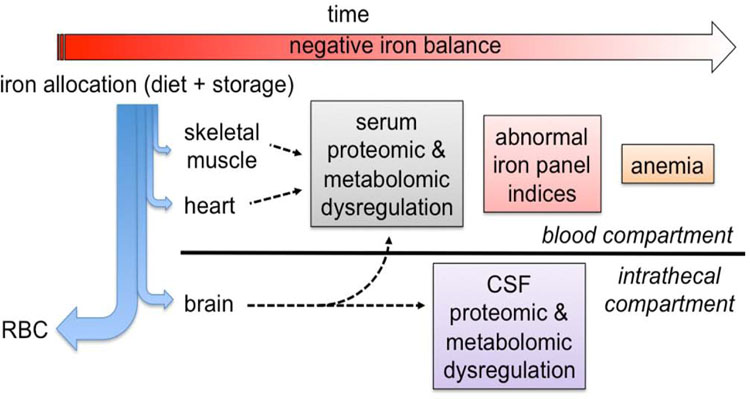
Figure 1: Iron prioritization during iron deficiency in fetal and neonatal sheep34,35,37 and monkeys.36.
The relative distributional flow of iron is indicated by the thickness of the blue arrows. The red blood cells receive the primary allocation followed sequentially by the brain, the heart and skeletal muscle.34–37 As negative iron balance progresses over time (red arrow), iron-dependent metabolic dysregulation of the skeletal muscle and heart is first noted by alterations in the serum metabolome36. Progressive worsening of iron deficiency subsequently negatively affects brain metabolism at approximately the same time that serum iron panels (eg, ferritin, %TSAT) become abnormal.36 Iron deficiency results in anemia only in the final stage of the process.34–37

